Home

Klem Paard Roeispaan ct scan t1 vs t2 Anekdote Over het algemeen heerlijkheid

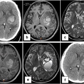
CT, T1 post-contrast, and T2 Flair images for illustrative Case 6. This...  | Download Scientific Diagram
CT, T1 post-contrast, and T2 Flair images for illustrative Case 6. This... | Download Scientific Diagram

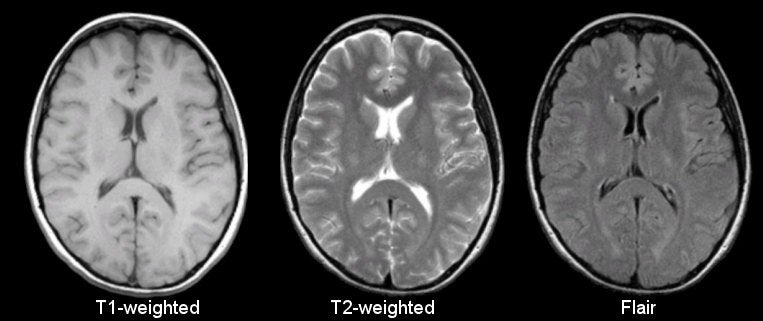
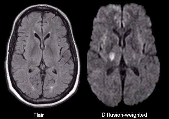
Comparison of Common MRI Sequences - T1 vs T2 vs Flair T1: ... | GrepMed

Advances in T1-weighted and T2-weighted imaging in the abdomen and pelvis.  | Semantic Scholar
Advances in T1-weighted and T2-weighted imaging in the abdomen and pelvis. | Semantic Scholar

Initial CT scan with left insular hematoma. b–d MRI-T1, T2, and T1... |  Download Scientific Diagram
Initial CT scan with left insular hematoma. b–d MRI-T1, T2, and T1... | Download Scientific Diagram

a) Axial CT, (b) axial T1-W, (c) T2-W and (d) diffusion-weighted MR... |  Download Scientific Diagram
a) Axial CT, (b) axial T1-W, (c) T2-W and (d) diffusion-weighted MR... | Download Scientific Diagram

MRI Basics
MRI Basics

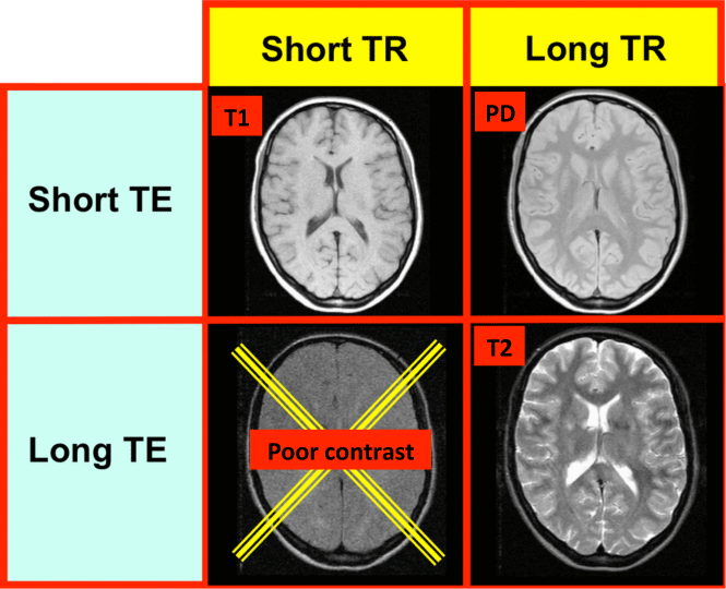
Image contrast - Questions and Answers ​in MRI

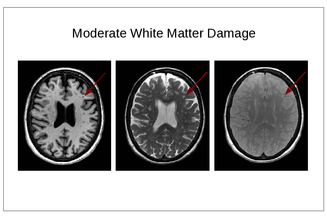
Four imaging modalities: (a) T1-weighted MRI; (b) T2-weighted MRI; (c)... |  Download Scientific Diagram
Four imaging modalities: (a) T1-weighted MRI; (b) T2-weighted MRI; (c)... | Download Scientific Diagram

A: MRI scan (T2 on the right and pre-contrast T1 on the left) showing a...  | Download Scientific Diagram
A: MRI scan (T2 on the right and pre-contrast T1 on the left) showing a... | Download Scientific Diagram

The Basics Left and Right The first step in reviewing radiology images is  knowing which side is left and which side is right. The images are  displayed in a standard fashion. When looking at a frontal image, the image  is oriented as if you are looking at the ...
The Basics Left and Right The first step in reviewing radiology images is knowing which side is left and which side is right. The images are displayed in a standard fashion. When looking at a frontal image, the image is oriented as if you are looking at the ...

Neuro-ophthalmology Question of the Week: Magnetic Resonance Imaging —  Neuro-Ophthalmology
Neuro-ophthalmology Question of the Week: Magnetic Resonance Imaging — Neuro-Ophthalmology

What is the difference between T1 and T2 imaging in MRI? - Quora

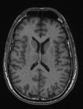
T1 MRI vs T2 MRI
T1 MRI vs T2 MRI

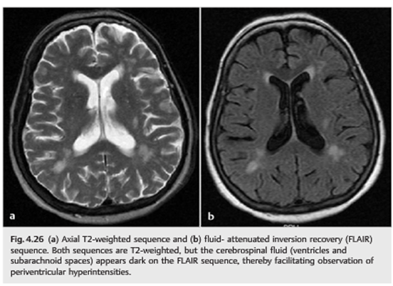
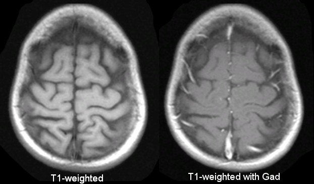
Brain Magnetic Resonance Imaging Technique: Approach Considerations, In the  MRI Machine, Guidelines
Brain Magnetic Resonance Imaging Technique: Approach Considerations, In the MRI Machine, Guidelines

MRI Basics
MRI Basics

Barone Mnemonic: MRI T1 verses T2

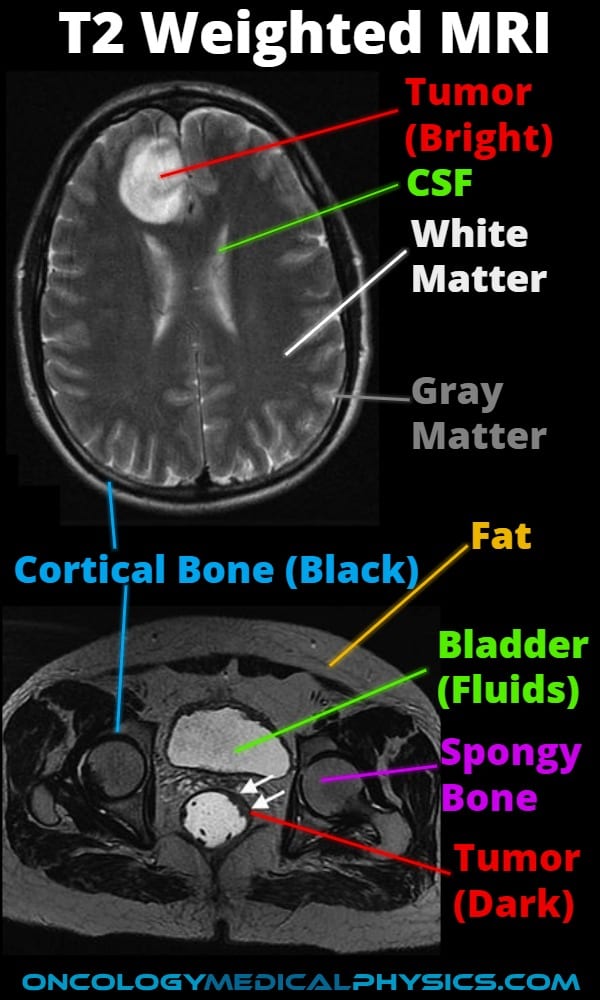
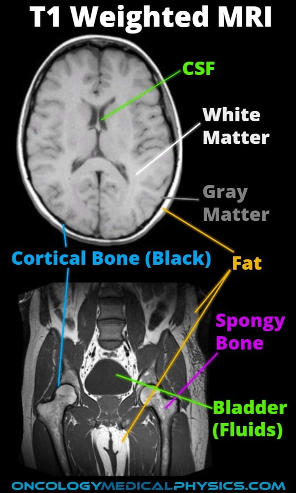
MRI Contrast Weighting | Oncology Medical Physics

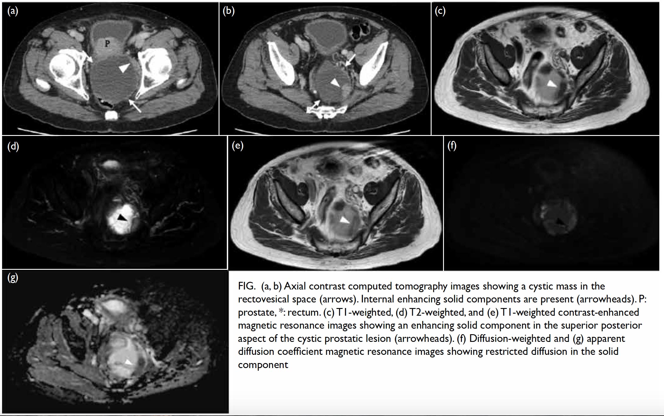
Cystic prostatic carcinoma: case report and literature review | HKMJ

3T How To: Structural MRI Imaging - Center for Functional MRI - UC San Diego

Contrast in MRI PD, T1, and T2. - ppt video online download

MRI Basics
MRI Basics

Figure 1. [CT and T1- and T2-weighted...]. - GeneReviews® - NCBI Bookshelf

Hong Kong College Of Radiologists: Answer of July 2016

Figure 2 from Synthetic vs. directly-acquired MRI of identical-slice brain  images: large scale and multi-contrast (PD, T1, and T2-weighted) image  quality comparison | Semantic Scholar
Figure 2 from Synthetic vs. directly-acquired MRI of identical-slice brain images: large scale and multi-contrast (PD, T1, and T2-weighted) image quality comparison | Semantic Scholar
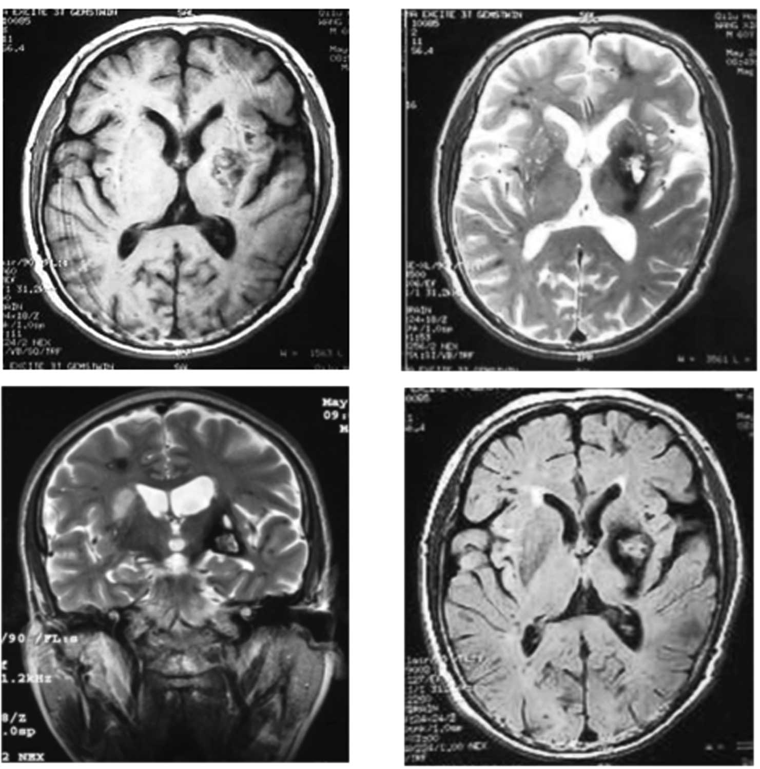
The value of T2*-weighted gradient echo imaging for detection of familial  cerebral cavernous malformation: A study of two families
The value of T2*-weighted gradient echo imaging for detection of familial cerebral cavernous malformation: A study of two families
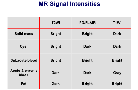
BASIC PRINCIPLES OF MR IMAGING John R. Hesselink, MD, FACR br-100.gif  Magnetic resonance (MR) is a dynamic and flexible technology that allows  one to tailor the imaging study to the anatomic part of interest and to the  disease process being studied ...
BASIC PRINCIPLES OF MR IMAGING John R. Hesselink, MD, FACR br-100.gif Magnetic resonance (MR) is a dynamic and flexible technology that allows one to tailor the imaging study to the anatomic part of interest and to the disease process being studied ...

MRI Contrast Weighting | Oncology Medical Physics

Classifications of atherosclerotic plaque components with T1 and T2*  mapping in 11.7 T MRI - ScienceDirect
Classifications of atherosclerotic plaque components with T1 and T2* mapping in 11.7 T MRI - ScienceDirect